Home

rovnat se rozsah Monarcha a431 cell line Kostýmy Rozsáhle neplacené

Authenticated A431 Cell Line Sigma Aldrich

Morphologies of infected A431 cell clones. Phase-contrast micrographs... |  Download Scientific Diagram
Morphologies of infected A431 cell clones. Phase-contrast micrographs... | Download Scientific Diagram

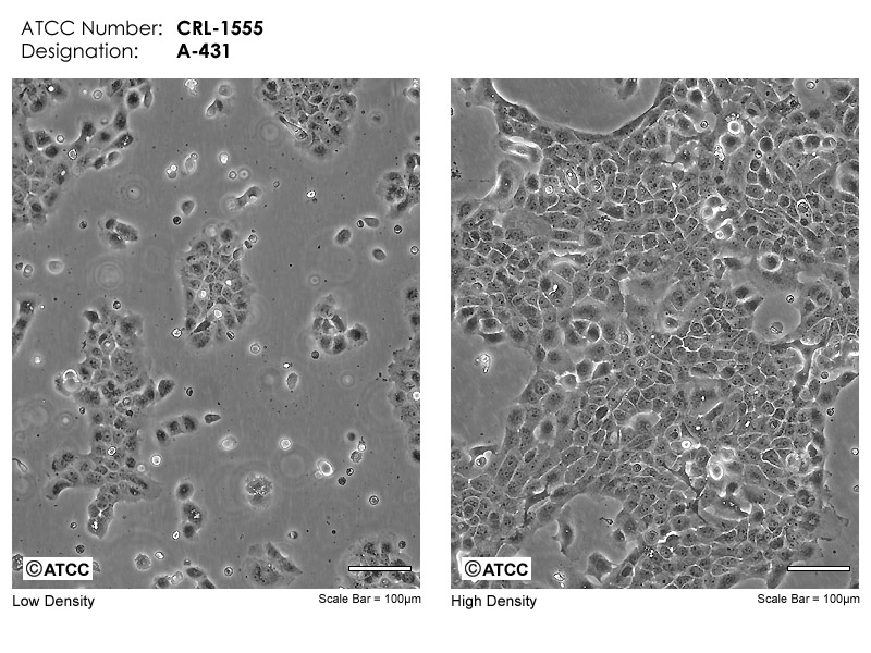
A-431 - CRL-1555 | ATCC

TTK Antibody (689913) (MAB6028): Novus Biologicals

Buy A431 | Cell Lines | CLS.shop

Microscopic images of A431 cells. The blue-colored areas are the cell... |  Download Scientific Diagram
Microscopic images of A431 cells. The blue-colored areas are the cell... | Download Scientific Diagram
At the Cell Counter: A431 Cells | Molecular Devices

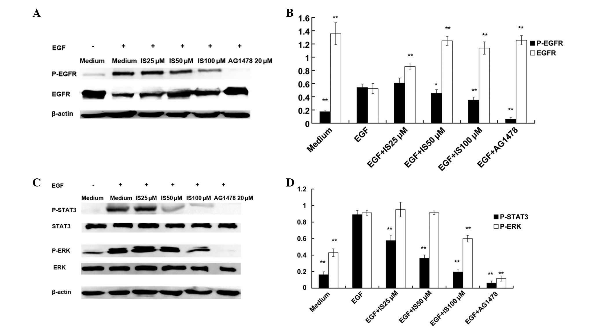
Icariside II induces apoptosis via inhibition of the EGFR pathways in A431  human epidermoid carcinoma cells
Icariside II induces apoptosis via inhibition of the EGFR pathways in A431 human epidermoid carcinoma cells
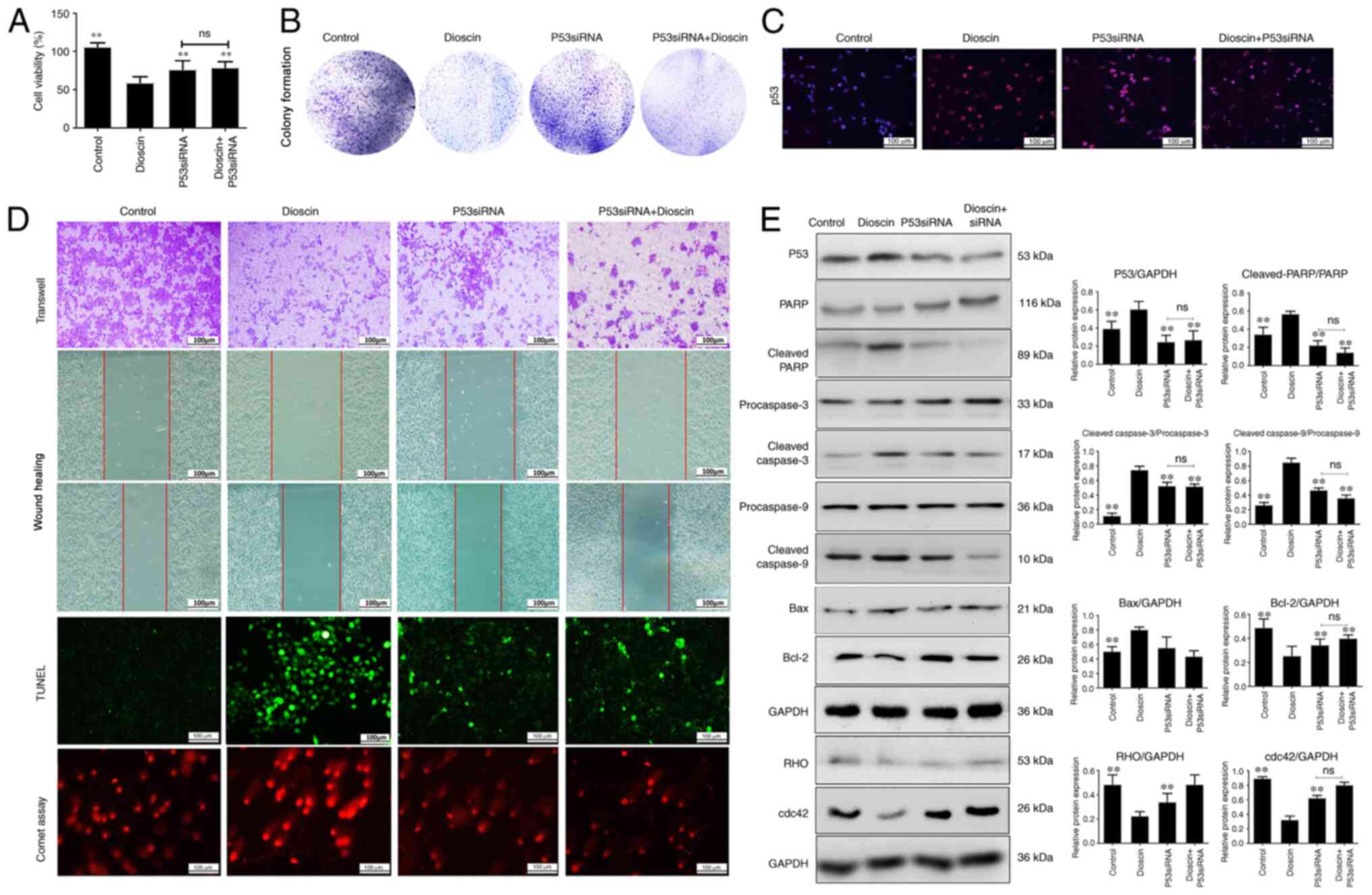
Antitumor effects of dioscin in A431 cells via adjusting ATM/p53‑mediated  cell apoptosis, DNA damage and migration
Antitumor effects of dioscin in A431 cells via adjusting ATM/p53‑mediated cell apoptosis, DNA damage and migration
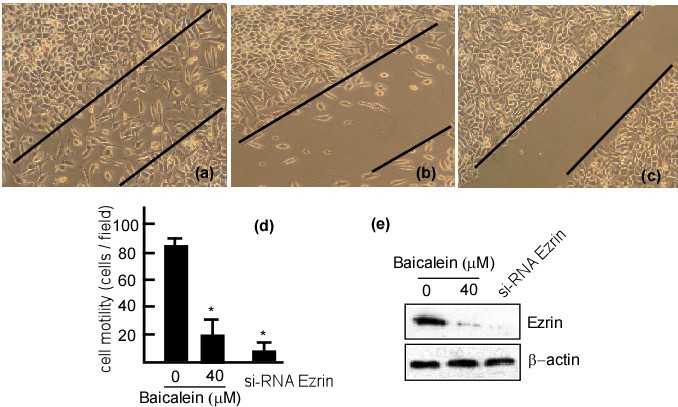
RETRACTED ARTICLE: Baicalein mediates inhibition of migration and  invasiveness of skin carcinoma through Ezrin in A431 cells | BMC Cancer |  Full Text
RETRACTED ARTICLE: Baicalein mediates inhibition of migration and invasiveness of skin carcinoma through Ezrin in A431 cells | BMC Cancer | Full Text

Expression of LMP2A and LMP2B in HaCat, SCC12F, and A431 epithelial... |  Download Scientific Diagram
Expression of LMP2A and LMP2B in HaCat, SCC12F, and A431 epithelial... | Download Scientific Diagram

PDF] Src induces morphological changes in A431 cells that resemble  epidermal differentiation through an SH3- and Ras-independent pathway. |  Semantic Scholar
PDF] Src induces morphological changes in A431 cells that resemble epidermal differentiation through an SH3- and Ras-independent pathway. | Semantic Scholar

A431D cells have fibroblast-like morphology. Living A431 cells (A and... |  Download Scientific Diagram
A431D cells have fibroblast-like morphology. Living A431 cells (A and... | Download Scientific Diagram
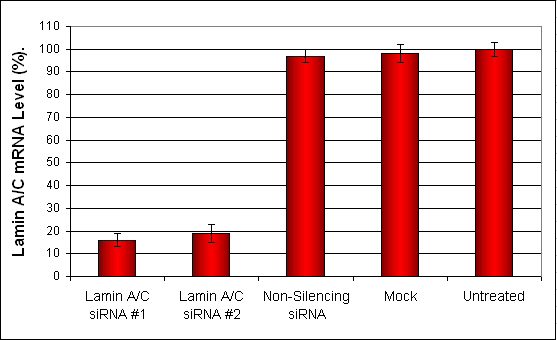
A431 Transfection Reagent (Epidermoid Carcinoma) | Transfection Reagents | Cell  Lines, In Vivo | Altogen Biosystems
A431 Transfection Reagent (Epidermoid Carcinoma) | Transfection Reagents | Cell Lines, In Vivo | Altogen Biosystems

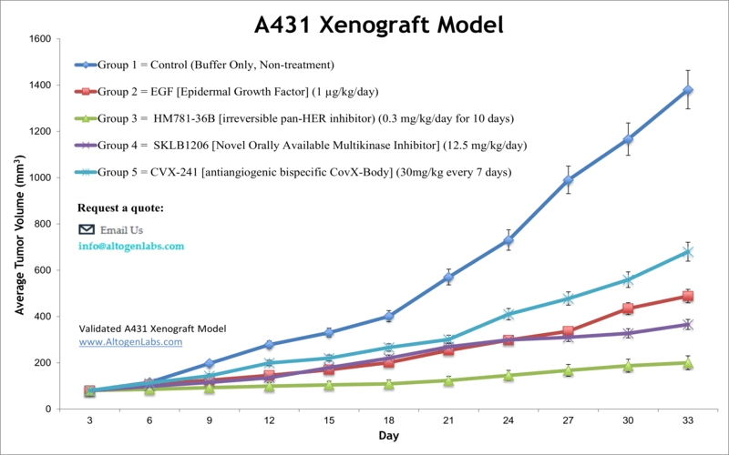
A431 Xenograft Model - Altogen Labs

Detailed Information [JCRB0004]-

Telomere shortening and apoptosis in telomerase-inhibited human tumor cells

Membrane Protein - Human Tumor Cell Line: A431 | BioChain Institute Inc.

Human KRT14 (Cytokeratin 14) knockout A-431 cell line (ab261897) | アブカム

Dasatinib worsens the effect of cetuximab in combination with fractionated  radiotherapy in FaDu- and A431-derived xenografted tumours | British  Journal of Cancer
Dasatinib worsens the effect of cetuximab in combination with fractionated radiotherapy in FaDu- and A431-derived xenografted tumours | British Journal of Cancer

Buy A431 (human epidermoid carcinoma) cell line slides | Axil Scientific

A431 from Cell Lines Service GmbH | SelectScience

GFP-Expressing Human A431 Epidermoid Carcinoma Cells-Angio-Proteomie

Identification of a Population of Epidermal Squamous Cell Carcinoma Cells  with Enhanced Potential for Tumor Formation | PLOS ONE
Identification of a Population of Epidermal Squamous Cell Carcinoma Cells with Enhanced Potential for Tumor Formation | PLOS ONE